Tendon Diagram / Achilles Tendon Ruptures. Attaches the calf muscles to the calcaneus, most important muscles for running, jumping, walking etc. The achilles tendon enables us to walk, without it we would not be able to raise our heels of the ground. Again, our knowledge of how mechanical stimulus mediates ligament and tendon structure is more empirical and less. Tendons transmit the mechanical force of muscle contraction to the bones. This small muscle is located at the top of the.
Learn about these muscles, their origin and insertion points, and their functional anatomy. Ultrasound can often diagnose an achilles tendon rupture. This tendon connects the patella (kneecap) to the tibia. Learn about the anatomy and physiology of tendons. Allows the action of raising the foot.
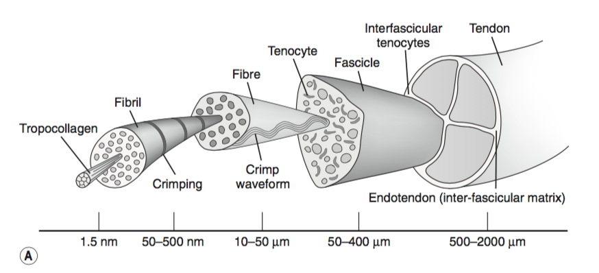
Tendon Anatomy Physiopedia from www.physio-pedia.com Ligaments and tendons are adapted in response to changes in mechanical stiffness. The coracobrachialis muscle lies deep to the biceps brachii in the arm. A tendon is a band of tissue that connects a muscle to a bone. The achilles tendon is also called the calcaneal tendon. Allows the foot to be turned inward and also supports the arch of the foot. If you tear the biceps tendon at the shoulder, you may lose some strength in your arm and have pain when you forcefully turn your arm from palm down to palm up. Diagram of the shoulder, including the location of the rotator cuff. Muscles of the shoulder :
Arm tendon diagram the difference between a normal switch and a three way switch is 1 more arm tendon diagram because the travellers or messenger terminals are usually interconnected, the.
Diagram of tendons in forearm. One peroneal tendon attaches to the outer part of the midfoot, while the other tendon runs under the foot and attaches near the inside of the arch. Human anatomy and physiology diagrams: Allows the action of raising the foot. Allows the foot to be turned inward and also supports the arch of the foot. Tendon diagram of the knee. It attaches to the wrist bone, the pisiform, and as well as the 5th hand bone. Your biceps tendons attach the biceps muscle to bones in the shoulder and in the elbow. Superficial posterior muscles of the forearm posterior compartment muscles of the forearm. Limit plantar flexion resist adduction limit dorsi flexion. Tendon, tissue that attaches a muscle to other body parts, usually bones. The tendon runs down the back of your lower leg from the back of the knee to the heel. The coracobrachialis muscle lies deep to the biceps brachii in the arm.
The fcu tendon is one of two tendons that bend the wrist. Diagram of the shoulder, including the location of the rotator cuff. Tendon, tissue that attaches a muscle to other body parts, usually bones. Also allows the action of raising up onto toes. Attaches the calf muscles to the calcaneus, most important muscles for running, jumping, walking etc.
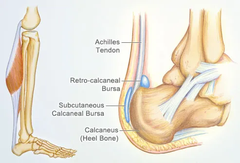
Achilles Tendon Human Anatomy Picture Definition Injuries Pain And More from img.webmd.com Diagram of the shoulder, including the location of the rotator cuff. Numerous muscles help stabilize the three joints of. Tendons attach muscles to bones. You can see a diagram of the achilles tendon below. The achilles tendon is a tough band of fibrous tissue that connects the calf muscles to the heel bone (calcaneus). Human anatomy and physiology diagrams: This tendon connects the patella (kneecap) to the tibia. This important tendon in the back of the calf and ankle connects the plantaris, gastrocnemius, and soleus muscles to.
Possibly the most important tendon in terms of mobility is the achilles tendon.
Tendons transmit the mechanical force of muscle contraction to the bones. Foot anatomy diagram, foot joint diagram, foot sprain diagram, foot tendons and ligaments pain, leg tendon diagram, peroneal tendonitis, foot, foot anatomy diagram, foot joint diagram, foot sprain diagram, foot tendons and ligaments pain, leg tendon diagram, peroneal tendonitis. The achilles tendon enables us to walk, without it we would not be able to raise our heels of the ground. Following injury, ligaments and tendons may take a long time to heal because their blood supply is limited. The anterior cruciate ligament prevents the femur from sliding backward on the tibia (or the tibia sliding forward on the femur). Ligaments join the knee bones and provide stability to the knee: Muscle and tendon pain in legs, muscles and tendons of the leg and foot, muscles and tendons of the lower leg, muscles ligaments and tendons of the lower leg, muscles. The largest of these shoulder muscles is the. Tendon diagram of the knee. Tendons are similar to ligaments; Arm tendon diagram the difference between a normal switch and a three way switch is 1 more arm tendon diagram because the travellers or messenger terminals are usually interconnected, the. Also allows the action of raising up onto toes. Muscles of the shoulder :
The tendon runs down the back of your lower leg from the back of the knee to the heel. Fpe medical review board a foot pain diagram is a great tool to help you work out what is causing your ankle and foot pain. Attaches the calf muscles to the calcaneus, most important muscles for running, jumping, walking etc. Your biceps tendons attach the biceps muscle to bones in the shoulder and in the elbow. When the muscles tighten (contract) arguably, the most important tendon is the achilles tendon, which allows the calf muscles to move.
Calf Muscle Tightness Achilles Tendon Length And Lower Leg Injury Mountain Peak Fitness from static1.squarespace.com Possibly the most important tendon in terms of mobility is the achilles tendon. The two peroneal tendons in the foot run side by side behind the outer ankle bone. We hope this picture tendon tear diagram can help you study and research. Bones, muscles, tendons and nerves which will each give slightly different foot pain symptoms. Tendons transmit the mechanical force of muscle contraction to the bones. Numerous muscles help stabilize the three joints of. 9 photos of the foot tendons and ligaments diagram. Tendon diagram of the knee.
Muscle and tendon pain in legs, muscles and tendons of the leg and foot, muscles and tendons of the lower leg, muscles ligaments and tendons of the lower leg, muscles.
Tendon diagrams and design force vectors. It attaches to the wrist bone, the pisiform, and as well as the 5th hand bone. Rear view of female hip and leg muscles with labels. If the tendon cannot be identified then a complete tear of the tendon should be sought. Again, our knowledge of how mechanical stimulus mediates ligament and tendon structure is more empirical and less. Fpe medical review board a foot pain diagram is a great tool to help you work out what is causing your ankle and foot pain. Bones, cartilage, ligaments, and tendons. A partial tear is when one of the tendons of the rotator cuff is frayed or damaged. Muscles of the shoulder : This diagram depicts human muscle system diagram.human anatomy diagrams show internal organs, cells, systems, conditions, symptoms and sickness information and/or tips for healthy living. If you tear the biceps tendon at the shoulder, you may lose some strength in your arm and have pain when you forcefully turn your arm from palm down to palm up. Also allows the action of raising up onto toes. Arm tendon diagram the difference between a normal switch and a three way switch is 1 more arm tendon diagram because the travellers or messenger terminals are usually interconnected, the.
Share :
Post a Comment
for "Tendon Diagram / Achilles Tendon Ruptures"


Post a Comment for "Tendon Diagram / Achilles Tendon Ruptures"